LEARN // THE TRANSPLANT ATLAS
The five organs that travel.
The liver, the kidney, the pancreas, the heart and the lungs: five organs that travel from a donor to a recipient. On each plate, read the donor's operation and then the recipient's: each step shades its part in red chalk (sanguine), and the joins appear as cut lines in the order they are usually made.
Drag to turn it; tap any part to name it
- PLATE VThe lungsOpen the plate
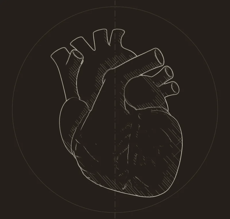
- PLATE IVThe heartOpen the plate
- PLATE IThe liverOpen the plate
- PLATE IIIThe pancreasOpen the plate
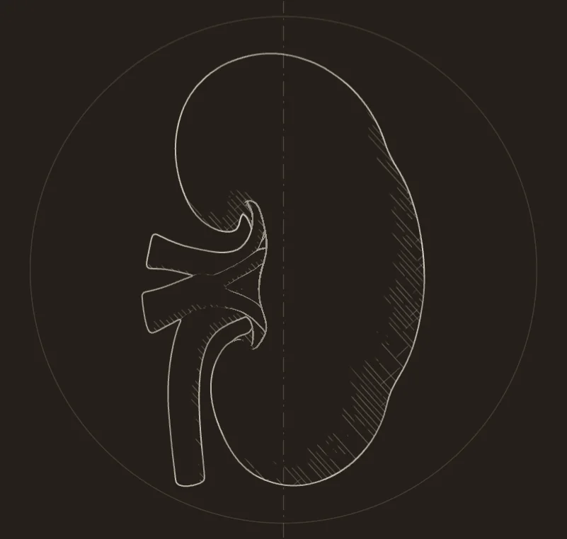
- PLATE IIThe kidneyOpen the plate
3D anatomy: BodyParts3D, © The Database Center for Life Science, licensed under CC Attribution-Share Alike 2.1 Japan. The models on this page are adapted from it and shared under the same licence.
LAYER 01 // THE SKIN
The surface comes first: the landmarks a surgeon feels before any incision, the clavicles, the sternal angle at the second rib, the costal margin and the iliac crests. Here the skin is a glass figure, so the body inside shows through.
LAYER 02 // THE MUSCLES
Beneath the skin, the superficial muscles: pectoralis major over the chest, the rectus abdominis down the front of the abdomen, the external oblique at its side, and the muscles of the limbs.
LAYER 03 // THE SKELETON
The frame that sets every organ at its level: twelve pairs of ribs, the vertebrae from the atlas to the fifth lumbar, the sacrum and the hip bones.
LAYER 04 // THE ORGANS AND THEIR VESSELS
Every organ in its place, with the aorta, the venae cavae and their branches. The five organs that travel are named in gold: choose one to open its plate.
- Skin
- Right sternocleidomastoid
- Left sternocleidomastoid
- Right trapezius
- Left trapezius
- Right deltoid
- Left deltoid
- Right pectoralis major
- Left pectoralis major
- Right serratus anterior
- Left serratus anterior
- Right latissimus dorsi
- Left latissimus dorsi
- Right external oblique
- Left external oblique
- Right rectus abdominis
- Left rectus abdominis
- Right biceps brachii
- Left biceps brachii
- Right triceps brachii
- Left triceps brachii
- Right brachioradialis
- Left brachioradialis
- Right forearm flexors
- Left forearm flexors
- Right gluteus maximus
- Left gluteus maximus
- Right gluteus medius
- Left gluteus medius
- Right tensor fasciae latae
- Left tensor fasciae latae
- Right sartorius
- Left sartorius
- Right rectus femoris
- Left rectus femoris
- Right vastus lateralis
- Left vastus lateralis
- Right vastus medialis
- Left vastus medialis
- Right adductor longus
- Left adductor longus
- Right gracilis
- Left gracilis
- Right tibialis anterior
- Left tibialis anterior
- Right fibularis longus
- Left fibularis longus
- Right gastrocnemius
- Left gastrocnemius
- Skull
- Mandible
- Hyoid bone
- Atlas (C1)
- Axis (C2)
- C3 vertebra
- C4 vertebra
- C5 vertebra
- C6 vertebra
- C7 vertebra
- T1 vertebra
- T2 vertebra
- T3 vertebra
- T4 vertebra
- T5 vertebra
- T6 vertebra
- T7 vertebra
- T8 vertebra
- T9 vertebra
- T10 vertebra
- T11 vertebra
- T12 vertebra
- L1 vertebra
- L2 vertebra
- L3 vertebra
- L4 vertebra
- L5 vertebra
- Intervertebral discs
- Right rib 1
- Right rib 2
- Right rib 3
- Right rib 4
- Right rib 5
- Right rib 6
- Right rib 7
- Right rib 8
- Right rib 9
- Right rib 10
- Right rib 11
- Right rib 12
- Left rib 1
- Left rib 2
- Left rib 3
- Left rib 4
- Left rib 5
- Left rib 6
- Left rib 7
- Left rib 8
- Left rib 9
- Left rib 10
- Left rib 11
- Left rib 12
- Right costal cartilage 1
- Right costal cartilage 2
- Right costal cartilage 3
- Right costal cartilage 4
- Right costal cartilage 5
- Right costal cartilage 6
- Right costal cartilage 7
- Right costal cartilages 8 to 10
- Left costal cartilage 1
- Left costal cartilage 2
- Left costal cartilage 3
- Left costal cartilage 4
- Left costal cartilage 5
- Left costal cartilage 6
- Left costal cartilage 7
- Left costal cartilages 8 to 10
- Manubrium of the sternum
- Body of the sternum
- Xiphoid process
- Sacrum
- Right clavicle
- Left clavicle
- Right scapula
- Left scapula
- Right humerus
- Left humerus
- Right radius
- Left radius
- Right ulna
- Left ulna
- Right hand bones
- Left hand bones
- Right hip bone
- Left hip bone
- Right femur
- Left femur
- Right patella
- Left patella
- Right tibia
- Left tibia
- Right fibula
- Left fibula
- Right foot bones
- Left foot bones
- Brain (the cerebral hemispheres)
- Cerebellum
- Brainstem
- Thymus
- Trachea
- Bronchial tree (main, lobar and segmental bronchi)
- Upper lobe of the right lung
- Middle lobe of the right lung
- Lower lobe of the right lung
- Upper lobe of the left lung
- Lower lobe of the left lung
- Heart
- Left coronary artery
- Right coronary artery
- Cardiac veins
- Gallbladder
- Stomach
- Spleen
- Duodenum
- Jejunum
- Ileum
- Colon
- Appendix
- Rectum
- Right adrenal gland
- Left adrenal gland
- Right ureter
- Left ureter
- Urinary bladder
- Oesophagus
- Diaphragm
- Liver
- Pancreas
- Right kidney
- Left kidney
- Ascending aorta
- Arch of the aorta
- Descending aorta (thoracic and abdominal)
- Brachiocephalic trunk
- Right common carotid artery
- Left common carotid artery
- Right subclavian artery
- Left subclavian artery
- Coeliac trunk
- Common hepatic artery
- Splenic artery
- Left gastric artery
- Superior mesenteric artery
- Inferior mesenteric artery
- Right renal artery
- Left renal artery
- Right renal vein
- Left renal vein
- Right common iliac artery
- Left common iliac artery
- Right external iliac artery
- Left external iliac artery
- Right internal iliac artery
- Left internal iliac artery
- Right common iliac vein
- Left common iliac vein
- Right external iliac vein
- Left external iliac vein
- Right internal iliac vein
- Left internal iliac vein
- Superior vena cava
- Inferior vena cava
- Right brachiocephalic vein
- Left brachiocephalic vein
- Right internal jugular vein
- Left internal jugular vein
- Right subclavian vein
- Left subclavian vein
- Splenic vein
- Superior mesenteric vein
- Pulmonary trunk and arteries
- Pulmonary veins
- Liver
- Gallbladder
- Inferior vena cava
- Coeliac trunk
- Common hepatic artery
- Splenic artery
- Left gastric artery
- Splenic vein
- Superior mesenteric vein
- Descending aorta (thoracic and abdominal)
- Stomach
- Spleen
- Pancreas
- Duodenum
- Right kidney
- Right adrenal gland
- Oesophagus
- Diaphragm
- Right renal artery
- Right renal vein
- Right rib 5
- Right rib 6
- Right rib 7
- Right rib 8
- Right rib 9
- Right rib 10
- Right rib 11
- Right rib 12
- Left rib 5
- Left rib 6
- Left rib 7
- Left rib 8
- Left rib 9
- Left rib 10
- Left rib 11
- Left rib 12
- Right costal cartilage 5
- Right costal cartilage 6
- Right costal cartilage 7
- Right costal cartilages 8 to 10
- Left costal cartilage 5
- Left costal cartilage 6
- Left costal cartilage 7
- Left costal cartilages 8 to 10
- Manubrium of the sternum
- Body of the sternum
- Xiphoid process
- T10 vertebra
- T11 vertebra
- T12 vertebra
- L1 vertebra
- L2 vertebra
- L3 vertebra
- Left kidney
- Right kidney
- Left adrenal gland
- Right adrenal gland
- Left ureter
- Right ureter
- Urinary bladder
- Right renal artery
- Left renal artery
- Right renal vein
- Left renal vein
- Descending aorta (thoracic and abdominal)
- Inferior vena cava
- Right common iliac artery
- Left common iliac artery
- Right external iliac artery
- Left external iliac artery
- Right internal iliac artery
- Left internal iliac artery
- Right common iliac vein
- Left common iliac vein
- Right external iliac vein
- Left external iliac vein
- Right internal iliac vein
- Left internal iliac vein
- Coeliac trunk
- Superior mesenteric artery
- Inferior mesenteric artery
- Right psoas major
- Left psoas major
- Right iliacus
- Left iliacus
- Right rib 11
- Right rib 12
- Left rib 11
- Left rib 12
- T11 vertebra
- T12 vertebra
- L1 vertebra
- L2 vertebra
- L3 vertebra
- L4 vertebra
- L5 vertebra
- Right hip bone
- Left hip bone
- Sacrum
- Pancreas
- Main pancreatic duct
- Duodenum
- Coeliac trunk
- Common hepatic artery
- Splenic artery
- Left gastric artery
- Superior mesenteric artery
- Inferior mesenteric artery
- Splenic vein
- Superior mesenteric vein
- Descending aorta (thoracic and abdominal)
- Inferior vena cava
- Left renal artery
- Left renal vein
- Right common iliac artery
- Left common iliac artery
- Right external iliac artery
- Left external iliac artery
- Right common iliac vein
- Left common iliac vein
- Right external iliac vein
- Left external iliac vein
- Liver
- Gallbladder
- Stomach
- Spleen
- Jejunum
- Right kidney
- Left kidney
- Urinary bladder
- T12 vertebra
- L1 vertebra
- L2 vertebra
- L3 vertebra
- L4 vertebra
- L5 vertebra
- Right hip bone
- Left hip bone
- Heart
- Aortic valve
- Mitral valve
- Tricuspid valve
- Pulmonary valve
- Papillary muscles of the left ventricle
- Papillary muscles of the right ventricle
- Left coronary artery
- Right coronary artery
- Cardiac veins
- Ascending aorta
- Arch of the aorta
- Descending aorta (thoracic and abdominal)
- Brachiocephalic trunk
- Right common carotid artery
- Left common carotid artery
- Right subclavian artery
- Left subclavian artery
- Pulmonary trunk and arteries
- Pulmonary veins
- Superior vena cava
- Inferior vena cava
- Right brachiocephalic vein
- Left brachiocephalic vein
- Upper lobe of the right lung
- Middle lobe of the right lung
- Lower lobe of the right lung
- Upper lobe of the left lung
- Lower lobe of the left lung
- Trachea
- Bronchial tree (main, lobar and segmental bronchi)
- Diaphragm
- Manubrium of the sternum
- Body of the sternum
- Xiphoid process
- Right rib 2
- Right rib 3
- Right rib 4
- Right rib 5
- Right rib 6
- Left rib 2
- Left rib 3
- Left rib 4
- Left rib 5
- Left rib 6
- Right costal cartilage 2
- Right costal cartilage 3
- Right costal cartilage 4
- Right costal cartilage 5
- Right costal cartilage 6
- Left costal cartilage 2
- Left costal cartilage 3
- Left costal cartilage 4
- Left costal cartilage 5
- Left costal cartilage 6
- Oesophagus
- Upper lobe of the right lung
- Middle lobe of the right lung
- Lower lobe of the right lung
- Upper lobe of the left lung
- Lower lobe of the left lung
- Trachea
- Bronchial tree (main, lobar and segmental bronchi)
- Pulmonary trunk and arteries
- Pulmonary veins
- Heart
- Ascending aorta
- Arch of the aorta
- Descending aorta (thoracic and abdominal)
- Superior vena cava
- Diaphragm
- Oesophagus
- Right rib 1
- Right rib 2
- Right rib 3
- Right rib 4
- Right rib 5
- Right rib 6
- Right rib 7
- Right rib 8
- Right rib 9
- Right rib 10
- Right rib 11
- Right rib 12
- Left rib 1
- Left rib 2
- Left rib 3
- Left rib 4
- Left rib 5
- Left rib 6
- Left rib 7
- Left rib 8
- Left rib 9
- Left rib 10
- Left rib 11
- Left rib 12
- Right costal cartilage 1
- Right costal cartilage 2
- Right costal cartilage 3
- Right costal cartilage 4
- Right costal cartilage 5
- Right costal cartilage 6
- Right costal cartilage 7
- Right costal cartilages 8 to 10
- Left costal cartilage 1
- Left costal cartilage 2
- Left costal cartilage 3
- Left costal cartilage 4
- Left costal cartilage 5
- Left costal cartilage 6
- Left costal cartilage 7
- Left costal cartilages 8 to 10
- Manubrium of the sternum
- Body of the sternum
- Xiphoid process
- Right clavicle
- Left clavicle
- T1 vertebra
- T2 vertebra
- T3 vertebra
- T4 vertebra
- T5 vertebra
- T6 vertebra
- T7 vertebra
- T8 vertebra
- T9 vertebra
- T10 vertebra
- T11 vertebra
- T12 vertebra
- Cantlie's line: right | left (schematic)
- Umbilical fissure (schematic)
- Hepatic artery proper (schematic)
- Portal vein (schematic)
- Hepatic veins into the IVC (schematic)
- Bile duct (schematic)
- Right lobe: segments V to VIII
- Left lobe: segments II to IV
- Left lateral section: II and III (schematic)
- Right lobe graft: the patient's right of Cantlie's line
- Right hepatic vein: one wide outflow
- Outflow: hepatic veins to the IVC
- Hepatic artery
- In front of the aorta, below the superior mesenteric artery
- Crosses in front of the aorta
- Superior mesenteric artery
- Artery stapled close to the aorta
- Vein divided
- Ureter clipped low
- Cold flush
- More than one artery is common
- Gibson incision
- Vein: end to side, external iliac vein
- Artery: end to side, external iliac artery
- Graft ureter (schematic)
- Ureter to the bladder dome (Lich-Gregoir)
- SMA stump
- Splenic artery stump
- Portal vein stump (schematic)
- Duodenum cut (first part)
- Duodenum cut (third part)
- Second part kept (papilla)
- Y-graft stem (schematic)
- External iliac limb to the SMA
- Internal iliac limb to the splenic artery
- Vein: end to side, distal IVC
- Artery: end to side, right common iliac artery
- Duodenum side to side to the small bowel
- The kidney: left iliac fossa, joins as Plate II
- Left atrial cuff
- IVC
- SVC
- Pulmonary artery
- Aorta
- Sinoatrial node (schematic)
- Cardioplegia
- Lung flush
- SVC ligated
- IVC clamped, cut above
- Aorta cross-clamped
- Left atrial appendage vented (schematic)
- Flush: main pulmonary artery
- Vent: left atrial appendage (schematic)
- Trachea stapled and cut
- Shortened to 1 or 2 rings above the lobar carina
- Clamshell: the 4th intercostal spaces
- Bronchus
- Atrial cuff
Scroll a plate's steps and each one washes its part. Tap a step, or a label on the plate, to show it.
FIG. 01 // LIVER TRANSPLANTATION
PLATE I // THE LIVER
A lobe, a section or the whole liver.
From the front, the falciform ligament divides the anatomical right and left lobes, but the functional division is Cantlie's line, from the gallbladder fundus to the inferior vena cava (IVC) in the plane of the middle hepatic vein2. The liver has eight Couinaud segments, each with its own portal triad branch, outflow and bile drainage, and the hepatic veins run between them2. A living donor gives a set of whole segments; a deceased donor's liver can go in whole3.
LAYER 01Anatomy
Learn the liver as a map of segments before you learn the operations: each of Couinaud's eight segments has its own independent inflow, outflow and biliary drainage, so the liver can be divided between segments into two working parts, a graft and the donor's remnant2.
The liver is the largest gland in the body and lies in the right upper quadrant beneath the right hemidiaphragm2. Its coronary ligament, the largest of its ligaments, leaves a bare area on its back, and the ligament's posterior layer attaches the liver to the right kidney2. At the hilum it is joined to the duodenum by the hepatoduodenal ligament, the right free edge of the lesser omentum, which carries the portal triad and forms the front wall of the epiploic (Winslow's) foramen2.
Source2
Cantlie's line runs from the gallbladder fundus to the suprahepatic inferior vena cava (IVC), in the plane of the middle hepatic vein, and divides the liver into right and left hemilivers; for decades the falciform ligament was believed to be the true division2. In the Brisbane 2000 terminology, segments II and III form the left lateral section and segment IV, divided into IVa above and IVb below, the left medial section; V and VIII form the right anterior section and VI and VII the right posterior section221. The portal branches run within the segments while the three major hepatic veins pass between them, and the caudate lobe, segment I, takes vessels and bile ducts from both the right and the left, and its veins drain directly into the IVC2.
In the usual pattern, seen in 757 of 1000 donor livers, the common hepatic artery arises from the coeliac axis, gives off the gastroduodenal artery and continues as the proper hepatic artery, which divides into right and left hepatic arteries9. The commonest variants were a replaced or accessory right hepatic artery from the superior mesenteric artery (SMA), in 106 livers, and a replaced or accessory left hepatic artery from the left gastric artery, in 97; in 15 the whole common hepatic artery came from the SMA9. The right hepatic artery passes behind the common hepatic duct in 80 to 90% of people2. Every variant artery has to be defined so that the whole graft is arterialised, and variant arterial anatomy is a risk factor for early hepatic artery thrombosis911.
The portal vein, about 8 cm long and without valves, begins where the splenic vein joins the superior mesenteric vein in front of the IVC; at the hilum its left branch has a longer, more horizontal course outside the liver, and its right branch divides into anterior and posterior branches2. Blood leaves by three major hepatic veins, right, middle and left: the middle and left share a trunk before the IVC in about 65 to 85% of people, and an accessory inferior right hepatic vein is present in 21 to 48%2. In a right-lobe graft, inferior right hepatic veins wider than 5 mm are joined to the IVC, and when the graft is taken without the middle hepatic vein, that vein's segment V and VIII branches wider than 5 mm are rebuilt, because undrained segments congest1417.
In the hepatoduodenal ligament the portal vein lies behind the common bile duct, which is slightly to its right, and the proper hepatic artery, which is slightly to its left2. The duct depends on the artery: hepatic artery occlusion is a cause of bile duct necrosis, and early non-anastomotic strictures are typically ischaemic-type biliary lesions13. Biliary complications are the most common complications after liver transplantation, with an incidence of 10 to 32%13.
Before a living donation the team estimates the graft's weight as a percentage of the recipient's body weight, the graft-to-recipient weight ratio (GRWR), and its volume against the recipient's standard liver volume; risk rises around a GRWR of 0.8% or a graft volume under 40% of the standard14. King Faisal Specialist Hospital and Research Centre (KFSHRC) offers its robotic recipient operation for right grafts under 900 g with an anticipated GRWR above 0.80%, and not when extra segment V or VIII veins would need joining4. After the transplant, Doppler ultrasound screens the hepatic artery, and magnetic resonance cholangiopancreatography (MRCP) is the gold standard for diagnosing biliary complications1113.
Pearls
- Cantlie's line, in the plane of the middle hepatic vein, divides the right and left hemilivers; the falciform ligament does not2.
- About one donor liver in four has a variant hepatic artery: in 1000 livers, 243 did not have the usual pattern9.
- A small graft is harmed by too much portal flow: excess flow raises sinusoidal pressure and, through the hepatic artery buffer response, lowers arterial flow14.
- Early hepatic artery thrombosis is less common in adults than in children, 2.9% against 8.3%, but about half of the affected grafts are replaced11.
- The caudate lobe, segment I, takes vessels from both the right and the left, and its veins drain directly into the IVC2.
LAYER 02The operation
The donor
Segments V to VIII, the usual adult living-donor graft. In the first three fully robotic cases at King Faisal Specialist Hospital and Research Centre (KFSHRC), Riyadh, the graft-to-recipient weight ratio was 0.81 to 1.10% (robotic recipients are chosen when it is expected to be above 0.80%), and the donors kept an estimated 32 to 42% of the liver4.
- Why
- An adult needs enough working liver. In 276 living-donor transplants, one-year actuarial graft survival in elective cases was 42% when the graft weighed less than 0.8% of the recipient's body weight, against 92% at 1 to 3%10, and a recent international consensus took a graft-to-recipient weight ratio (GRWR) of 0.8 as the safe limit for adults14. The right lobe is the usual adult graft: in one Taiwanese series of 380 donors, 93.4% gave a right hemi-liver16.
- Watch for
- Small-for-size syndrome, with cholestasis, coagulopathy and ascites, develops when a graft is too small for its portal flow and for the recipient's needs; the excess flow injures the sinusoids14. Segment V and VIII veins are branches of the middle hepatic vein: when the graft is taken without that vein, branches over 5 mm are rebuilt with grafts and smaller ones tied17.
- On the plate
- The right lobe washes: segments V to VIII, everything to the right of Cantlie's line, which on this front view is on your left.
Segments II to IV. KFSHRC has reported its first two fully robotic left-graft living donor liver transplants6.
- Why
- A left lobe leaves the donor more of the liver: in that series, left-lobe donors kept a larger share of their total liver volume than right-lobe donors16. Recent studies have not shown an adverse impact of using left lobe grafts14.
- Watch for
- Its weak point is size: a left lobe graft may contain less functioning liver than a right lobe graft of the same volume14, and grafts under 0.8% of the recipient's weight did worse10.
- On the plate
- The left lobe washes: segments II to IV, to the left of Cantlie's line, which on this view is on your right.
Segments II and III, the left lateral section21.
- Why
- Segments II and III form one section, and each segment has its own inflow, outflow and biliary drainage, so the section can be taken whole221. It is used for children, and for a small infant even this can be too big: it can be reduced further, into a hyperreduced graft, to prevent large-for-size complications15.
- Watch for
- Too large is a risk as well as too small: in Kiuchi's series, grafts of 3% or more of the recipient's weight had more vascular complications and acute rejection10. Children's arteries clot more often: early hepatic artery thrombosis was 8.3% in children against 2.9% in adults11.
- On the plate
- The left lateral section washes, the far left of the liver, which on this view is the tip on the right of the plate.
The donor operation divides the liver along the planes drawn on the plate, so that each graft is a set of whole segments. KFSHRC reported in 2024 that it offers robotic donor hepatectomy to all its donors, after more than 1000 minimally invasive donor hepatectomies4.
- Why
- Each segment has its own independent inflow, outflow and biliary drainage, and the major hepatic veins run between segments, so a cut along those planes leaves both the graft and the donor's remnant working2. The remnant then regrows: in 380 donors it regenerated to a volume similar to the whole liver before donation16.
- Watch for
- Donor safety comes first. A 2026 meta-analysis of 7 studies with more than 15,000 adult donors found fewer transfusions and less overall donor morbidity with robotic than with laparoscopic donor hepatectomy, and no difference in major complications or deaths18. The learning curve is steep, because liver tissue is fragile and bleeding needs experienced control19.
- On the plate
- The division planes appear as cut lines: one along Cantlie's line between the right and left lobes, and one between segment IV and the left lateral section.
The back table
On the back table, a right lobe taken with its middle hepatic vein has that vein joined to the right hepatic vein, to make one wide triangular opening17. In a right lobe taken without that vein, segment V and VIII branches over 5 mm are rebuilt with expanded polytetrafluoroethylene (ePTFE) grafts, which are then joined to the right hepatic vein as one opening, and smaller ones are tied; in both, inferior right hepatic veins over 5 mm are kept for their own join to the IVC17.
- Why
- Every part of the graft must drain: reconstruction of the hepatic venous outflow is essential to prevent graft congestion17, and undrained segments also impair the liver beside them14.
- Watch for
- The width of the opening matters: in that series of 52 grafts, an anastomotic diameter of 30 mm or less raised the risk of outflow complications (odds ratio 14.3), and one occlusion was fatal17. Each join was checked by Doppler ultrasound on the back table and again after reperfusion17.
- On the plate
- The right lobe washes again: its outflow is built at its top edge, where the hepatic veins leave it for the IVC.
The recipient
A whole liver goes in by piggyback, which keeps the recipient's IVC and needs less bypass and transfusion, or by the conventional technique, which replaces the recipient's IVC and traditionally uses veno-venous bypass, a pump that carries blood from the legs and gut back to the heart while the IVC is clamped3. For a right-lobe living-donor graft, its right hepatic vein is joined to the IVC under a partial clamp4.
- Why
- The outflow is joined first, before any blood is let into the graft4. A graft that cannot drain congests, and undrained areas also impair the liver around them14. Piggyback keeps the recipient's IVC: in a network meta-analysis of 40 studies and 10,238 patients, piggyback methods shortened the operation and needed fewer transfusions than caval replacement, with similar results afterwards12.
- Watch for
- A kinked or narrow outflow obstructs the graft. In a series of 1067 transplants, the graft's upper cava was kept short to prevent kinking and the recipient's three hepatic veins were opened into one cuff to widen the join; venous outflow stenosis followed 0.54% of the piggyback implantations3. Caval replacement was still chosen when a tumour lay close to the IVC3.
- On the plate
- The whole graft washes, and a cut line runs along its top, where the hepatic veins meet the IVC under the diaphragm.
Joined second4. In the hepatoduodenal ligament the portal vein lies behind, with the common bile duct to its right and the hepatic artery to its left12.
- Why
- With the outflow ready, the portal vein is joined next, with a continuous 6/0 suture in the robotic series4, and releasing the portal clamp reperfuses the graft20.
- Watch for
- Reperfusion can cause post-reperfusion syndrome: the blood pressure falls and the pulmonary artery pressure goes up, thought to be from the cold, acidic blood that enters the circulation when the clamp is released20. In a small living-donor graft the danger is too much portal flow; a portal pressure under 15 mmHg is recommended, particularly with an older donor or a marginal graft14.
- On the plate
- The cut line across the portal vein, the middle of the three structures below the liver.
Joined third, with interrupted sutures in the robotic series4.
- Why
- Without arterial flow the bile duct can die: hepatic artery occlusion is a cause of bile duct necrosis13. Early hepatic artery thrombosis complicated 4.4% of 21,822 transplants in a systematic review; 53.1% of those grafts were replaced and a third of the patients died11.
- Watch for
- Reported risk factors include arterial conduits, variant arterial anatomy, a long operation and low recipient weight11. High portal flow can also lower arterial flow, through the hepatic artery buffer response14. The robotic series uses fine interrupted sutures, 7/0 or 8/04.
- On the plate
- The cut line on the vessel below the liver to the right of the portal vein on the plate (the patient's left): the hepatic artery.
Last: duct-to-duct4, or a Roux-en-Y hepaticojejunostomy, to a loop of jejunum. In primary sclerosing cholangitis, a 2026 meta-analysis of 12 non-randomised studies linked Roux-en-Y with better patient and graft survival, though duct-to-duct caused less cholangitis8.
- Why
- The bile duct comes last4. Duct-to-duct keeps the sphincter of Oddi and endoscopic access, takes less time and carries a lower risk of septic complications; a loop of jejunum is used when the recipient has biliary disease, the ducts differ in size, or after earlier biliary surgery or retransplantation13.
- Watch for
- Leaks come early, and strictures early or late: leaks are about 23% of biliary complications and anastomotic strictures about 47%13. Strictures follow 5 to 15% of transplants, and up to 30% after living-donor grafts13.
- On the plate
- The cut line on the bile duct, the structure to the left of the portal vein on the plate (the patient's right).
Robotic
At KFSHRC the donor hepatectomy and all four recipient joins have been done by robot4567; the native liver comes out, and the graft goes in, through a 12-cm lower abdominal incision4. In August 2023 the centre reported what it describes as the world's first fully robotic recipient hepatectomy and graft implantation, with a graft from a fully robotic living donor4; a first series of 10 followed5, then left grafts6 and children: its first series of purely robotic liver transplants in children had 7, 6 partial grafts done fully by robot and 1 whole liver from a domino donor implanted by robot7.
Where practice varies
Practice varies: a whole liver goes in by piggyback or with caval replacement3, and the bile duct is joined duct-to-duct or to a Roux-en-Y loop8.
Sources 21
- Gray H. Anatomy of the Human Body. 20th ed. 1918 (the archive.org scan), p. 681. Open the source (opens in a new tab)
- Kostov S, Selçuk I, Watrowski R, et al. Surgical anatomy of the liver: significance in ovarian cancer surgery. Diagnostics 2023;13(14):2371. doi:10.3390/diagnostics13142371 (opens in a new tab)
- Nishida S, Nakamura N, Vaidya A, et al. Piggyback technique in adult orthotopic liver transplantation: 1067 liver transplants. HPB 2006;8(3):182-8. doi:10.1080/13651820500542135 (opens in a new tab)
- Broering DC, Raptis DA, Elsheikh Y. Pioneering fully robotic donor hepatectomy and robotic recipient liver graft implantation: a new horizon in liver transplantation. Int J Surg 2024;110(3):1333-1336. doi:10.1097/JS9.0000000000001031 (opens in a new tab)
- Broering DC, Elsheikh Y, Malago M, et al. Outcomes of fully robotic recipient living donor liver transplant in relation to the open approach. Transplantation 2024;108(12):2396-2402. doi:10.1097/TP.0000000000005109 (opens in a new tab)
- Broering DC, Raptis DA, Elsheikh Y. Fully robotic recipient left graft living donor liver transplantation. Am J Transplant 2025;25(8):1784-1787. doi:10.1016/j.ajt.2025.03.027 (opens in a new tab)
- Broering DC, Rescigno L, Elsheikh Y, et al. First series of purely robotic liver transplantation in children. Am J Transplant 2026. doi:10.1016/j.ajt.2026.10.007 (opens in a new tab)
- Faleiro MD, et al. Duct-to-duct versus hepaticojejunostomy reconstruction after liver transplantation for primary sclerosing cholangitis: meta-analysis. Transplantation 2026;110(4):e774-e784. doi:10.1097/TP.0000000000005639 (opens in a new tab)
- Paper Hiatt JR, Gabbay J, Busuttil RW. Surgical anatomy of the hepatic arteries in 1000 cases. Ann Surg 1994;220(1):50-2. doi:10.1097/00000658-199407000-00008 (opens in a new tab) PubMed 8024358 (opens in a new tab) Open the source (opens in a new tab)
- Paper Kiuchi T, Kasahara M, Uryuhara K, et al. Impact of graft size mismatching on graft prognosis in liver transplantation from living donors. Transplantation 1999;67(2):321-7. doi:10.1097/00007890-199901270-00024 (opens in a new tab) PubMed 10075602 (opens in a new tab)
- Paper Bekker J, Ploem S, de Jong KP. Early hepatic artery thrombosis after liver transplantation: a systematic review of the incidence, outcome and risk factors. Am J Transplant 2009;9(4):746-57. doi:10.1111/j.1600-6143.2008.02541.x (opens in a new tab) PubMed 19298450 (opens in a new tab)
- Paper Khajeh E, Ramouz A, Aminizadeh E, et al. Comparison of the modified piggyback with standard piggyback and conventional orthotopic liver transplantation techniques: a network meta-analysis. HPB (Oxford) 2023;25(7):732-746. doi:10.1016/j.hpb.2023.02.017 (opens in a new tab) PubMed 37120378 (opens in a new tab)
- Paper Vernuccio F, Mercante I, Tong XX, et al. Biliary complications after liver transplantation: a computed tomography and magnetic resonance imaging pictorial review. World J Gastroenterol 2023;29(21):3257-3268. doi:10.3748/wjg.v29.i21.3257 (opens in a new tab) PubMed 37377585 (opens in a new tab) Open the source (opens in a new tab)
- Paper Reddy MS, Gopal PV. Small for size syndrome in living donor liver transplantation: prevention and management. J Clin Exp Hepatol 2025;15(2):102458. doi:10.1016/j.jceh.2024.102458 (opens in a new tab) PubMed 39722782 (opens in a new tab) Open the source (opens in a new tab)
- Paper Namgoong JM, Hwang S, Song GW, et al. Pediatric liver transplantation with hyperreduced left lateral segment graft. Ann Hepatobiliary Pancreat Surg 2020;24(4):503-512. doi:10.14701/ahbps.2020.24.4.503 (opens in a new tab) PubMed 33234754 (opens in a new tab) Open the source (opens in a new tab)
- Paper Wang WC, Wu TH, Hung HC, et al. Liver regeneration of living donor after liver donation for transplantation: disparity in the left and right remnant liver. Medicine (Baltimore) 2024;103(14):e37632. doi:10.1097/MD.0000000000037632 (opens in a new tab) PubMed 38579088 (opens in a new tab) Open the source (opens in a new tab)
- Paper Le HT, Vu QV, Le TV, Tran TM. Long-term patency and complications of single orifice hepatic venous outflow reconstruction in right lobe graft living donor liver transplantation: a Vietnamese center experience. Ann Hepatobiliary Pancreat Surg 2025;29(4):390-395. doi:10.14701/ahbps.25-109 (opens in a new tab) PubMed 40825725 (opens in a new tab) Open the source (opens in a new tab)
- Paper Al Sayed M, Albashier M, Altabbaa H, et al. From laparoscopy to robotics in living donor hepatectomy: a systematic review and meta-analysis of comparative outcomes. J Robot Surg 2026;20(1). doi:10.1007/s11701-026-03360-2 (opens in a new tab) PubMed 42133269 (opens in a new tab) Open the source (opens in a new tab)
- Paper Kakos CD, Papanikolaou A, Ziogas IA, Tsoulfas G. Global dissemination of minimally invasive living donor hepatectomy: what are the barriers? World J Gastrointest Surg 2023;15(5):776-787. doi:10.4240/wjgs.v15.i5.776 (opens in a new tab) PubMed 37342850 (opens in a new tab) Open the source (opens in a new tab)
- Paper Gao Q, Cai JZ, Dong H. A review of the risk factors and approaches to prevention of post-reperfusion syndrome during liver transplantation. Organogenesis 2024;20(1):2386730. doi:10.1080/15476278.2024.2386730 (opens in a new tab) PubMed 39097866 (opens in a new tab) Open the source (opens in a new tab)
- Paper Agostini A, Borgheresi A, Floridi C, et al. The role of imaging in surgical planning for liver resection: what the radiologist need to know. Acta Biomed 2020;91(8-S):18-26. doi:10.23750/abm.v91i8-S.9938 (opens in a new tab) PubMed 32945275 (opens in a new tab) Open the source (opens in a new tab)
FIG. 02 // KIDNEY TRANSPLANTATION
PLATE II // THE KIDNEY
Into the right iliac fossa.
The plate looks into the recipient's right iliac fossa, where most kidney grafts sit, outside the peritoneum24. The graft is a living donor's left kidney turned over, so its pelvis and ureter lie in front and its vein deepest11516; the small figure at the top right shows that kidney in the donor. The native kidneys are usually left in place: even in polycystic disease, only 18.7% of recipients at one centre had one or both removed14.
LAYER 01Anatomy
Two operations in two places: the donor's kidney comes out from behind the peritoneum, and the graft goes into the recipient's iliac fossa, outside the peritoneum, beside the psoas muscle915. Two pieces of anatomy recur through the steps: the order of the structures at the hilum, and the fact that the graft ureter has only one blood supply15.
In the donor the left kidney lies behind the peritoneum: it is reached by moving the left colon towards the midline to open the retroperitoneal space, and then opening Gerota's fascia9. In the recipient the graft goes into the iliac fossa by an extraperitoneal approach, in a gutter made beside the psoas muscle15. The right fossa is usually preferred, because its external iliac vein lies more superficially3 and runs a more horizontal course15.
At the hilum the structures keep a stable order from front to back: the renal vein, then the artery, then the pelvis and ureter15. When a kidney goes into the opposite side's fossa, as a left kidney into the right, its collecting system becomes the most anterior structure, which keeps the ureter away from the iliac vessels15. That is why the graft on this plate shows its pelvis and ureter in front and its vein deepest15.
Source15
Multiple renal arteries are common: they were seen in 18.2% of 214 living donors in one series, without worse graft function10. Not every artery has to be saved: one under 1 mm, especially to the upper pole, may be tied if the area left without blood is under 15% of the upper or middle pole, but lower-pole arteries over 1 mm should be joined if possible, to prevent ischaemia of the ureter3. The graft ureter relies solely on blood from the preserved renal artery branches15.
The Kidney Disease: Improving Global Outcomes (KDIGO) guideline advises taking the left kidney when there is no reason to take the right, because its longer venous pedicle makes the operation easier18. The left renal vein's tributaries, the gonadal, adrenal and lumbar veins, are divided in the donor operation9. In a meta-analysis of 105 studies, a retroaortic left renal vein, running behind the aorta, was found in about 3% of people, a circumaortic one, ringing the aorta, in about 3.5%, and multiple renal veins in 16.7%, far more often on the right (16.6%) than on the left (2.1%)19.
The transplanted ureter relies solely on blood from the preserved renal artery branches, which run in the tissue around it; ischaemia of its distal end is a leading cause of early ureteric problems, and most leaks occur at that distal end15. So the ureter is taken with at least 1 cm of that tissue, and cut as short as possible to reduce later ischaemic complications3.
KDIGO recommends renal imaging, for example computed tomography (CT) angiography, in every living donor candidate18; it maps the number, course and branching of the renal vessels, especially the renal vein10. When the two kidneys differ, or one has an abnormality that still allows donation, the more severely affected kidney is the one donated18. After transplantation, vascular complications occur in 3 to 15% of recipients, and image-guided treatment is preferred to surgical revision20.
Pearls
- Front to back at the hilum: vein, artery, pelvis; a left kidney placed in the right fossa is turned so that its pelvis comes to the front15.
- The left kidney is taken unless there is a reason to take the right: its longer vein makes the operation easier18.
- No non-transfixing clip on a donor's renal artery: KDIGO says such clips should not be used there, and calls for a suture ligature or a staple anchored in the vessel wall18.
- The graft ureter has one blood supply, the renal artery branches, so it is kept short and in its own sleeve of tissue315.
LAYER 02The operation
The donor
Laparoscopic living-donor nephrectomy is the gold standard28. The donor keeps the better kidney; if both are equal the left is taken8, because its renal vein is longer710.
- Why
- Donor safety decides first: when the kidneys differ in function, or one has an abnormality that does not prevent donation, the Kidney Disease: Improving Global Outcomes (KDIGO) guideline says the more severely affected kidney should be donated, so the donor keeps the better one18. Otherwise the left kidney is taken in laparoscopic donor nephrectomy, because its longer venous pedicle makes the technique easier18.
- Watch for
- The anatomy must be known before the operation: KDIGO recommends renal imaging, for example computed tomography (CT) angiography, in every donor candidate18. It maps the number, course and branching of the vessels, especially the renal vein, and multiple renal arteries are common: 18.2% of 214 donors in one series10.
- On the plate
- The small framed figure at the top right washes: the donor's left kidney in place, before any cut.
It crosses in front of the aorta just below the superior mesenteric artery, with the left renal artery behind it1. The right kidney can be taken too, its vein sometimes extended with gonadal or iliac vein10.
- Why
- Its length is why the left kidney is preferred18; the operation frees it by dividing the veins that join it, the gonadal, adrenal and lumbar veins, before the renal vein itself is stapled beyond the adrenal vein9.
- Watch for
- Check the CT for a variant: a retroaortic left renal vein is found in about 3% of people and a circumaortic one in about 3.5%19. A right kidney has its own trap: multiple renal veins are much more common on the right, 16.6% against 2.1% on the left19.
- On the plate
- In the donor figure: the long left renal vein crossing in front of the aorta, the adrenal vein entering from above, the gonadal vein from below and a lumbar vein behind (dashed); the artery runs hidden behind the vein.
The gonadal and adrenal veins are sealed and divided and the lumbar vein clipped. The ureter is clipped as low as possible and divided, then the renal artery is stapled close to the aorta and the renal vein beyond the adrenal vein, and both are divided9. In one study of male donors, clipping the ureter and gonadal vein above where they cross the iliac vessels prevented orchialgia, pain in the testis11.
- Why
- Everything is ready before the blood supply is cut: in the procedure outline the back table is set up, the ureter divided and a retrieval bag placed first; then the artery and vein are stapled, and the kidney is removed at once and flushed with cold preservation solution9. One series of 63 donors prepared the extraction site at the start to reduce the warm ischaemia time10.
- Watch for
- Secure the artery by transfixion: KDIGO says non-transfixing clips such as Hem-o-lok should not be used on the donor's renal artery, only a suture ligature or a staple anchored in the vessel wall18, after reports of harm that included a donor's death10. Keep the tissue around the ureter, at least 1 cm of it3, because the ureter's blood vessels run through it15.
- On the plate
- The donor's cut lines appear: the artery and the vein at the aorta, the artery's cut behind the vein's, and the ureter low.
The back table
On the back table the graft's blood is washed out and replaced with cold preservation solution, a mandatory step3. Small accessory veins and the major lumbar veins are suture-ligated, the ureter keeps at least 1 cm of its surrounding tissue, and removing the fat around the kidney can wait until after revascularisation3.
- Why
- Each tie has a purpose: the small veins are suture-ligated to prevent bleeding from the hilum after the operation, and some surgeons ligate all the major lymphatic vessels to prevent a later lymphocele3; the tissue around the ureter is kept because the ureter's blood vessels run through it15.
- Watch for
- Check the vessel lengths for the side chosen: a right renal artery or a left renal vein that is too long will kink, and a short vein from a deceased donor can be lengthened with a piece of that donor's external iliac vein, internal jugular vein or inferior vena cava3.
- On the plate
- The graft washes on its own, before any join is made.
Multiple renal arteries are common, 18.2% of 214 living donors in one series10. A deceased donor's arteries can stay together on one Carrel patch of aorta; otherwise two arteries of similar size are joined side to side, or a small one end-to-side into a larger one315.
- Why
- Joined on the bench, several arteries can go into the recipient through one arterial join315. Not every artery must be saved: one under 1 mm, especially at the upper pole, may be tied if less than 15% of the upper or middle pole is left without blood3.
- Watch for
- Lower-pole arteries over 1 mm should be joined if possible, to prevent ischaemia of the ureter3, which relies solely on the preserved renal artery branches for its blood15.
- On the plate
- The graft washes again: its arteries are prepared before the arterial join is made.
The recipient
Open surgery uses a curved Gibson incision in the lower quadrant, parallel to the inguinal ligament from the anterior superior iliac spine to the midline 2 cm above the pubis, and the graft goes outside the peritoneum2; the hockey-stick incision is a common variant23. The right side is usually preferred because its external iliac vein is more superficial3.
- Why
- The right iliac fossa is usually preferred because its external iliac vein lies more superficially3 and runs a more horizontal course, which makes the venous join simpler15. In the open operation the inferior epigastric vessels, and in women the round ligament, are usually tied and divided, while the spermatic cord is simply retracted medially3.
- Watch for
- Lymphocele, a collection of lymph beside the graft, is the trap here: the lymphatics around the iliac vein are tied as it is prepared, and avoiding excessive dissection is the most important prevention3. Joining the artery to the internal iliac artery slightly increases it3.
- On the plate
- The fossa washes, with both cut lines: the curved Gibson incision low on the abdomen and the short midline incision used for the robot.
Outside the peritoneum in the right iliac fossa24, in the gutter beside the psoas muscle15, so it lies lateral to the iliac vessels with its hilum turned towards them. A left kidney is turned front to back to face the right iliac vessels, which brings its pelvis and ureter to the front11516.
- Why
- The graft is laid in a gutter made beside the psoas muscle, outside the peritoneum, with its hilum towards the iliac vessels15. Turning a left kidney over brings its collecting system to the front, which keeps the ureter away from the iliac vessels15.
- Watch for
- Vessel length must suit the position: a right renal artery or a left renal vein that is too long will kink, causing graft dysfunction and hypertension3.
- On the plate
- The graft washes, lateral to the iliac vessels, with its pelvis and ureter in front and its vein the deepest structure.
The renal vein goes end-to-side to the external iliac vein215, first when the artery goes end-to-side to the external iliac artery15. On this plate it passes deep to the external iliac artery to reach the vein.
- Why
- When the artery will go end-to-side to the external iliac artery, the vein is joined first, so that the arterial join can then be aligned; when the artery goes to the internal iliac artery, the artery goes first, which helps position the vein15.
- Watch for
- Thrombosis of the graft's artery or vein occurs in 0.1 to 8.2% of transplants20. A long clamp on the iliac vein adds a risk of deep vein thrombosis, which is why one technique chapter joins the artery first, to shorten it3.
- On the plate
- The cut line where the renal vein meets the external iliac vein, lower and more medial than the arterial join; the renal vein passes deep to the artery to reach it.
End-to-side to the external iliac artery, with a Carrel patch of aorta when the kidney comes from a deceased donor, or end-to-end to the internal iliac artery, when the artery may go first2315.
- Why
- End-to-side to the external iliac artery is generally preferred, especially when the donor artery carries a healthy Carrel patch of aorta; the internal iliac artery is more prone to atherosclerosis15. Several arteries can be brought together on one Carrel patch, joined separately, or joined side to side first15.
- Watch for
- Arterial stenosis is the commonest vascular complication, in 3 to 12.5% of recipients20. A polar artery that is tied leaves part of the kidney without blood, so lower-pole arteries over 1 mm should be joined if possible3.
- On the plate
- The cut line where the renal artery meets the external iliac artery, above the venous join.
Once blood flows through the kidney, the ureter is joined to the bladder dome or side wall, not the trigone, where the native ureters enter, by an extravesical Lich-Gregoir ureteroneocystostomy: the bladder mucosa is opened under the reflected detrusor, the bladder muscle, and the ureter sewn to it235; the detrusor is then closed over it to make a short anti-reflux tunnel3. It leaks less and causes less haematuria than the Politano-Leadbetter technique5, and a routine stent probably reduces major urological complications6.
- Why
- The order of unclamping varies: one technique chapter releases the artery first and the veins once the graft has filled3, while a pictorial essay removes the venous clamp first15. The ureter is then cut to the smallest possible length, to reduce later ischaemic complications, and joined without tension315; every technique aims to prevent leakage, stricture and reflux3.
- Watch for
- Most leaks occur at the distal end of the graft ureter, where ischaemia is a leading cause of early problems15. Stents are debated because of the risk of infection; one centre uses one and removes it after 3 weeks3.
- On the plate
- The ureter and the bladder wash, with the cut line where the ureter enters the upper side wall of the bladder.
Robotic
In robot-assisted kidney transplantation the robot makes every join, vein, artery and ureter, with the same technique as open surgery; the first fully robotic transplant, in Chicago, was done through a 7-cm midline incision2. The Vattikuti-Medanta technique places the ports as for a robotic prostatectomy and cools the graft with ice slush through a gel-sealed access port12. One concern when the graft lies inside the peritoneum is that it may twist2. Robotic donor nephrectomy, first reported in 2002, is safe, with results similar to laparoscopy10. The KDIGO 2017 living-donor guideline, citing limited experience and safety data at the time, advised that robotic donor nephrectomy should generally not be used18. KFSHRC reports more than 3000 minimally invasive transplant procedures across kidney and liver, donors and recipients17.
Where practice varies
Vessel choice varies: in one large series of 1,262 deceased-donor transplants, joining the vein of a left kidney placed on the right to the inferior vena cava (IVC) or the common iliac vein shortened the operation by a median of 17 minutes13, and the artery may go end-to-end to the internal iliac artery315. The fossa's cut line shows both incisions, the Gibson curve and a short midline incision for the robot, whose exact site varies by team2.
Sources 20
- Gray H. Anatomy of the Human Body. 20th ed. 1918 (the archive.org scan), pp. 602, 610, 613-614, 622, 672, 677, 679, 1223, 1227, 1233-1234. Open the source (opens in a new tab)
- Spiers HVM, Sharma V, Woywodt A, et al. Robot-assisted kidney transplantation: an update. Clin Kidney J 2022;15(4):635-643. doi:10.1093/ckj/sfab214 (opens in a new tab)
- Kakaei F, Nikeghbalian S, Malekhosseini SA. Kidney transplantation techniques. In: Rath T, ed. Current Issues and Future Direction in Kidney Transplantation. IntechOpen; 2013. doi:10.5772/54829 (opens in a new tab)
- Bickle I, Hacking C, Sechenov N, et al. Renal transplant. Reference article, Radiopaedia.org (last revised 30 June 2026). doi:10.53347/rID-44860 (opens in a new tab)
- Alberts VP, Idu MM, Legemate DA, et al. Ureterovesical anastomotic techniques for kidney transplantation: a systematic review and meta-analysis. Transpl Int 2014;27(6):593-605. doi:10.1111/tri.12301 (opens in a new tab)
- Patterson LG, Tingle SJ, Rix DA, et al. Routine intraoperative ureteric stenting for kidney transplant recipients. Cochrane Database Syst Rev 2024;7:CD004925. doi:10.1002/14651858.CD004925.pub4 (opens in a new tab)
- Wang K, Zhang P, Xu X, Fan M. Right versus left laparoscopic living-donor nephrectomy: a meta-analysis. Exp Clin Transplant 2015;13(3):214-26. Open the source (opens in a new tab)
- Musquera Felip M, Peri Cusí L, Alcaraz Asensio A. Surgical aspects of living-donor kidney transplantation. Nefrologia 2010;30 Suppl 2:71-9 (in Spanish). Open the source (opens in a new tab)
- Kimura S, Kawai T. Left laparoscopic donor nephrectomy (procedure outline). J Med Insight 2024. doi:10.24296/jomi/170 (opens in a new tab)
- Patel R, Fleetwood VA, Bhusal S, et al. Non-invasive approaches to living donor nephrectomy. Curr Transplant Rep 2025;12. doi:10.1007/s40472-025-00487-3 (opens in a new tab)
- Sureka SK, Srivastava A, Agarwal S, et al. Prevention of orchialgia after left-sided laparoscopic donor nephrectomy. J Endourol 2015;29(6):696-9. doi:10.1089/end.2014.0645 (opens in a new tab)
- Menon M, Sood A, Bhandari M, et al. Robotic kidney transplantation with regional hypothermia (IDEAL phase 2a). Eur Urol 2014;65(5):991-1000. doi:10.1016/j.eururo.2013.12.006 (opens in a new tab)
- Emmanouilidis N, Hashem AAB, Stiegler P, et al. Transplanting a left or right donor kidney into the left or right iliac fossa. Updates Surg 2023;75(5):1243-1257. doi:10.1007/s13304-023-01512-9 (opens in a new tab)
- Fuller TF, Brennan TV, Feng S, et al. End stage polycystic kidney disease: indications and timing of native nephrectomy. J Urol 2005;174(6):2284-8. doi:10.1097/01.ju.0000181208.06507.aa (opens in a new tab)
- Novacescu D, Latcu SC, Raica M, et al. Surgical strategies for renal transplantation: a pictorial essay. J Clin Med 2024;13(14):4188. doi:10.3390/jcm13144188 (opens in a new tab)
- Starzl TE, Marchioro TL, Morgan WW, Waddell WR. A technique for use of adult renal homografts in children. Surg Gynecol Obstet 1964;119:106-8. Open the source (opens in a new tab)
- Broering DC, Raptis DA, Malago M, Clavien PA, et al. Revolutionizing organ transplantation with robotic surgery. Ann Surg 2024;280(5):706-712. doi:10.1097/SLA.0000000000006460 (opens in a new tab)
- Guideline Kidney Disease: Improving Global Outcomes (KDIGO) Living Kidney Donor Work Group (Lentine KL, Kasiske BL, Levey AS, et al.). KDIGO Clinical Practice Guideline on the Evaluation and Care of Living Kidney Donors. Transplantation 2017;101(8S Suppl 1):S1-S109. Recommendations 5.9, 17.1, 17.4, 17.5, 17.6. doi:10.1097/TP.0000000000001769 (opens in a new tab) PubMed 28742762 (opens in a new tab) Open the source (opens in a new tab)
- Paper Hostiuc S, Rusu MC, Negoi I, Dorobanțu B, Grigoriu M. Anatomical variants of renal veins: a meta-analysis of prevalence. Sci Rep 2019;9(1):10802. doi:10.1038/s41598-019-47280-8 (opens in a new tab) PubMed 31346244 (opens in a new tab) Open the source (opens in a new tab)
- Paper Verloh N, Doppler M, Hagar MT, et al. Interventional management of vascular complications after renal transplantation. Rofo 2023;195(6):495-504. doi:10.1055/a-2007-9649 (opens in a new tab) PubMed 36863368 (opens in a new tab)
FIG. 03 // PANCREAS TRANSPLANTATION
PLATE III // THE PANCREAS
With its own duodenum.
The graft is a whole pancreas from a deceased donor, usually taken in a multi-organ retrieval, with a short segment of the donor's duodenum3. The plate shows the common open layout of a simultaneous pancreas-kidney transplant (SPK): a midline laparotomy, both grafts inside the peritoneum, the pancreas on the right with its head up, and the kidney in the left iliac fossa234. At KFSHRC the first simultaneous kidney-pancreas transplant was done in 20047, and a series of 27 consecutive pancreas transplants, 10 SPK and 17 pancreas after kidney, has been reported8.
LAYER 01Anatomy
The pancreas graft comes with more than the gland: a segment of the donor's duodenum, into which its main duct opens and which carries its exocrine juice into the recipient's bowel or bladder310, and two separate arteries that are joined into one before implantation25.
The pancreas lies at the level of the transpyloric plane (L1) and, apart from its tail, behind the peritoneum10. Its head sits in the C-shaped curve of the duodenum, with the aorta and the inferior vena cava (IVC) behind it; the stomach lies in front, across the lesser sac; and the tail, the only intraperitoneal part, reaches the splenic hilum in the splenorenal ligament with the splenic vessels10.
Source10
From right to left come the head, with the hook-like uncinate process below it lying behind the superior mesenteric vessels; the neck, which overlies those vessels; the body, which crosses the midline behind the stomach; and the tail10. The main pancreatic duct runs the length of the gland and joins the common bile duct to form the ampulla, which opens at the major duodenal papilla in the second, descending part of the duodenum, surrounded by the sphincter of Oddi110.
The head, uncinate process and duodenum are supplied through the pancreaticoduodenal arcade: the superior pancreaticoduodenal artery, from the gastroduodenal artery, and the inferior pancreaticoduodenal artery, from the superior mesenteric artery (SMA)510. The body and tail are supplied mainly by branches of the splenic artery5. A graft therefore keeps both the SMA and the splenic artery, and the Y-graft turns them into a single arterial inflow25.
The head drains into the superior mesenteric vein (SMV) and the body and tail into the splenic vein, both tributaries of the portal vein510. The graft's portal vein is cut above the duodenum, leaving enough length for the venous join5; if it is still too short, a donor iliac vein extends it2.
After the transplant, colour and spectral Doppler ultrasound is essential for checking the graft's vessels, multiphase computed tomography (CT) angiography with late arterial and venous phases shows them in detail, and magnetic resonance imaging (MRI) gives better contrast resolution5. The main threat is clotting: acute thrombosis of a graft vessel occurs in 2 to 10% of recipients, most often in the vein and usually within 4 to 6 weeks5.
Source5
A simultaneous pancreas-kidney transplant (SPK) is for people with type 1 diabetes and uraemia, a pancreas after kidney (PAK) for those whose kidney transplant already works, and a pancreas transplant alone (PTA) for those without uraemia3; the 2021 World Consensus found that SPK also benefits suitable recipients with type 2 diabetes, improving survival and quality of life compared with staying on dialysis9. It also found that SPK and pancreas alone can improve long-term survival, and that every type improves quality of life9.
Pearls
- Two arteries in, one vein out: the SMA and the splenic artery come in through the Y-graft, and the portal vein drains the graft25.
- Y-graft limbs: the external iliac limb goes to the SMA and the internal iliac limb to the splenic artery5.
- Clots are the commonest technical cause of early loss: vascular thrombosis made up 50% of technical failures in the first three months3.
- Neither venous route wins: portal drainage does not clearly improve metabolic parameters, and systemic drainage is technically more accessible59.
LAYER 02The operation
The donor
The pancreas comes out with the duodenum and spleen, keeping stumps of the superior mesenteric artery (SMA), the splenic artery and the portal vein3. The gastroduodenal artery (GDA) is ligated, and the SMA beyond the inferior pancreaticoduodenal artery; the superior mesenteric vein (SMV) below the pancreas and the splenic vessels at the splenic hilum are tied, and the portal vein is cut above the duodenum5.
- Why
- The pancreas is removed after the liver, en bloc with the duodenum and spleen, keeping its vessel stumps for the back table3; the SMA is divided at its origin from the aorta and the portal vein above the duodenum, leaving enough length for the venous join5. The donor matters too: risk factors for surgical complications include a donor older than 45, a body mass index over 28 and death from cerebrovascular disease3.
- Watch for
- A damaged gland is discarded: signs of acute pancreatitis, oedema, haematoma or fatty infiltration lead to discard3. Long storage harms it too: preservation for more than 20 hours is a risk factor for complications3.
- On the plate
- The whole graft washes: head, neck, body and tail, with the duodenal segment at the top.
On the back table, the side table where the graft is prepared in ice-cold solution, the spleen is removed and the duodenum shortened, keeping only its second part, stapled23. That segment carries the papilla, where the main pancreatic duct opens1.
- Why
- The duodenum is shortened with staplers, keeping only the second part, so that the enteric drainage has no redundant bowel2. That segment carries the major papilla, where the main duct opens, so it becomes the graft's exocrine outlet into the recipient's bowel or bladder310.
- Watch for
- The duodenal segment is where leaks start: intestinal leaks follow 5 to 8% of transplants, mostly in the immediate postoperative period3. On the back table its ends are closed and invaginated3.
- On the plate
- Only the duodenal segment washes, the short loop at the top of the graft, before any join is made.
The donor's common iliac artery with its fork: the external iliac limb is joined to the SMA and the internal iliac limb to the splenic artery5, so one arterial join to the recipient is enough2. A donor iliac vein extension is used if the portal vein is short2.
- Why
- The graft has two arteries, the SMA and the splenic artery; the Y-graft gives them a single arterial inflow, which simplifies the reconstruction and minimises the warm ischaemia time2.
- Watch for
- Match the limbs: the external iliac limb goes to the SMA and the internal iliac limb to the splenic artery5. Arterial thrombosis makes up about a quarter to a third of graft thromboses, and it comes early5.
- On the plate
- The Y-graft washes with its two bench joins, to the SMA and to the splenic artery; the stem's join to the recipient comes later.
The back table
If the graft's portal vein is not long enough, a donor iliac vein is added to give a tension-free venous join2. A final flush with methylene blue then shows any bleeding points before the graft goes in2.
- Why
- The aim is a tension-free venous join2, because venous thrombosis is the commonest vascular complication of the graft5.
- Watch for
- Bleeding and pseudoaneurysm are among the most life-threatening complications, at about 2.4%5; the methylene blue flush finds sources of bleeding while they can still be dealt with on the back table2.
- On the plate
- The whole graft washes on the back table, before any join is made.
The recipient
Vein before artery2. The graft's portal vein goes end-to-side to the inferior vena cava (IVC) or an iliac vein, systemic drainage, as drawn here45.
- Why
- The vein is joined before the artery2. Systemic drainage into the iliac vein or IVC is technically more accessible and so is preferred, while portal drainage is more physiological; neither shows a clear difference in glucose control or graft survival5, and the 2021 World Consensus found that portal drainage does not clearly improve metabolic parameters9.
- Watch for
- Venous thrombosis is the commonest vascular complication, usually within 4 to 6 weeks5, and splenic or portal vein thrombosis is the leading cause of early graft loss2. In the robotic technique the head-down position gives the portal vein a straight course to the external iliac vein, for free outflow without stasis2.
- On the plate
- The cut line on the graft's portal vein, where it meets the recipient's IVC behind the graft.
The stem of the Y-graft goes end-to-side to the recipient's right common or external iliac artery5.
- Why
- With the bench work done, one join is enough: the Y-graft's common iliac stem goes end-to-side to the recipient's iliac artery5.
- Watch for
- Arterial pseudoaneurysm and bleeding are among the most life-threatening complications, at about 2.4%5. Arterial thrombosis is an early complication, so the arterial flow is checked with Doppler ultrasound5.
- On the plate
- The cut line where the stem of the Y-graft meets the recipient's right common iliac artery, below the venous join.
Source5
Exocrine drainage is enteric in over 90%: a side-to-side duodeno-jejunostomy or duodeno-ileostomy, or a Roux-en-Y loop of jejunum245. Bladder drainage, a duodenocystostomy, is still used, mainly in pancreas-alone transplants, where urinary amylase helps to monitor rejection45; it can cause metabolic acidosis, from bicarbonate lost in the urine, dehydration, haematuria and reflux pancreatitis34, and 10 to 25% of patients, up to half in some series, later need conversion to enteric drainage35.
- Why
- Enteric drainage is used in over 90% of transplants5. Bladder drainage is not associated with more immediate surgical complications, but it carries more urological and metabolic complications9, which lead to enteric conversion in up to 50% of cases5.
- Watch for
- The exocrine leak is an early complication, reported in up to 20% of pancreas transplants, though it causes less than 1% of graft losses5. Graft pancreatitis is among the most common complications, and early acute pancreatitis raises the risk of graft loss at one year5.
- On the plate
- The duodenal segment washes with its cut line, the side-to-side join to a loop of the recipient's small bowel at the top of the plate.
At the same operation the kidney usually goes in the opposite iliac fossa, here the left, with the same joins as on Plate II2.
- Why
- A simultaneous pancreas-kidney transplant is for type 1 diabetes with uraemia3, and the World Consensus found that it can improve long-term survival, also in suitable recipients with type 2 diabetes9. In the open operation the two grafts usually go on opposite sides, the pancreas on the right and the kidney on the left2.
- Watch for
- The pancreas may lie inside the peritoneum: on low-quality data, the World Consensus found no evidence that intraperitoneal placement increases surgical complications9. The kidney's joins are those of Plate II2.
- On the plate
- The kidney washes at the lower right of the plate, in the left iliac fossa, with its vessels running out of view.
Robotic
A reproducible fully robotic technique has been described in a paper co-authored by Prof. Dieter Broering2: the pancreas goes on the left, head down and outside the peritoneum, through a lower abdominal incision. The portal vein goes end-to-side to the external iliac vein, then the Y-graft to the external iliac artery, and the bowel join is a side-to-side duodeno-ileostomy, with the kidney of an SPK on the right. The robot makes every join; the Y-graft is made by hand on the back table before2. The 2021 World Consensus judged robotic pancreas transplantation feasible, but found the data too limited to conclude on its safety9.
Where practice varies
Practice varies between centres: the side and direction of the graft, systemic or portal venous drainage, and enteric or bladder drainage245. Neither venous drainage has clear metabolic or survival advantages, and systemic drainage is simpler and more common45; portal drainage goes to the SMV, for example behind the right colon6. In open SPK the pancreas usually goes on the right and the kidney on the left; robotic practice usually reverses this2.
Sources 10
- Gray H. Anatomy of the Human Body. 20th ed. 1918 (the archive.org scan), pp. 605, 607, 681-682, 1208-1210. Open the source (opens in a new tab)
- Abreu P, Kadri H, Zaragoza S, et al. (with Broering D, Boggi U, Spaggiari M). The rebirth of pancreas transplantation: an innovative, fully robotic, reproducible surgical technique. BMC Surg 2026;26(1). doi:10.1186/s12893-026-03860-w (opens in a new tab)
- Meirelles Júnior RF, Salvalaggio P, Pacheco-Silva A. Pancreas transplantation: review. Einstein (São Paulo) 2015;13(2):305-9. doi:10.1590/S1679-45082015RW3163 (opens in a new tab)
- D'Alessandro C, Todisco M, Di Bella C, et al. Surgical complications after pancreatic transplantation: a CT pictorial review. World J Gastroenterol 2023;29(46):6049-6059. doi:10.3748/wjg.v29.i46.6049 (opens in a new tab)
- Torikai H, Swensson J, Fromke E, et al. Multimodality diagnostic imaging and role of interventional radiology in pancreas transplantation. Abdom Radiol 2025;50(12):5782-5798. doi:10.1007/s00261-025-05003-w (opens in a new tab)
- Boggi U, Vistoli F, Signori S, et al. A technique for retroperitoneal pancreas transplantation with portal-enteric drainage. Transplantation 2005;79(9):1137-42. doi:10.1097/01.tp.0000157279.39761.cc (opens in a new tab)
- Al Meshari K. The kidney transplant program at King Faisal Specialist Hospital and Research Center. Saudi J Kidney Dis Transpl 2005;16(4):586-97. Open the source (opens in a new tab)
- Gunther Brockmann J, Butt A, AlHussaini HF, et al. Protocol duodenal graft biopsies aid pancreas graft surveillance. Transplantation 2019;103(3):622-629. doi:10.1097/TP.0000000000002412 (opens in a new tab)
- Guideline Boggi U, Vistoli F, Andres A, et al. First World Consensus Conference on pancreas transplantation: Part II - recommendations. Am J Transplant 2021;21 Suppl 3:17-59. Statement 1.6, recommendations 5.1, 5.2, 5.7 and 5.10, jury deliberation A.3, and the conclusions in its abstract. doi:10.1111/ajt.16750 (opens in a new tab) PubMed 34245223 (opens in a new tab) Open the source (opens in a new tab)
- Reference TeachMeAnatomy. The pancreas (online anatomy reference, last updated 6 November 2025). Open the source (opens in a new tab)
FIG. 04 // HEART TRANSPLANTATION
PLATE IV // THE HEART
Five joins, the left atrium first.
From the front, the right ventricle forms most of the surface and the left ventricle the left border; of the left atrium only the auricle shows1. Near the heart the pulmonary trunk lies in front of the aorta1. The bicaval transplant joins the heart in five places, and the first, the left atrium, lies at the back: on the plate it is the loop behind the base of the heart2.
LAYER 01Anatomy
A heart transplant replaces the pump but keeps the recipient's back wall: the recipient keeps a cuff of posterior left atrium around its own pulmonary veins, and the donor heart is joined to that cuff, to the two venae cavae and to the two great arteries25.
The heart lies in the middle mediastinum, inside the pericardium6. Its anterior (sternocostal) surface is formed by the right ventricle, its left border by the left ventricle with part of the left atrium, its right border by the right atrium, and its base, at the back, by the left atrium6. Behind the ascending aorta and pulmonary trunk, and in front of the superior vena cava (SVC), the transverse pericardial sinus separates the arterial vessels from the venous ones6.
Source6
A bicaval transplant makes five joins: the left atrium, the inferior vena cava (IVC), the SVC, the pulmonary artery and the aorta2. The deepest is the left atrium, at the base of the heart: the recipient keeps a cuff of posterior left atrium around the entry of its pulmonary veins, and the donor's cuff is made by connecting the incisions between its four pulmonary veins56. The recipient's SVC is cut at the cavo-atrial junction, and a wide IVC cuff is left5.
The coronary arteries run in the sulci: the right coronary artery and the circumflex branch of the left coronary artery in the coronary (atrioventricular) sulcus with the coronary sinus, the anterior interventricular (left anterior descending) artery with the great cardiac vein on the front, and the posterior interventricular artery with the middle cardiac vein behind6. A donor heart is checked for coronary disease: coronary angiography is advised in donors with risk factors such as age over 45 or diabetes5.
The sinoatrial node lies in the upper wall of the right atrium, where the SVC enters, and the atrioventricular node in the atrioventricular septum near the opening of the coronary sinus7. The biatrial technique's right atrial incision can cause rhythm disorders: in one series sinus rhythm was present at discharge in only about 82% of biatrial recipients2.
The phrenic nerves, the only motor nerves of the diaphragm, run on the fibrous pericardium: the right lateral to the SVC and over the right atrium, the left over the left ventricle, each in front of its lung root9. Injury to one paralyses its hemidiaphragm, which then moves paradoxically, and injury to both can be life-threatening9. The left recurrent laryngeal nerve loops around the aortic arch just beyond the ligamentum arteriosum, and injury to one recurrent nerve gives a hoarse, breathy voice10.
A persistent left SVC occurs in about 0.3% of people and in up to 5% of those with congenital abnormalities8. In a transplant recipient it should be dealt with early in the operation, to keep venous drainage uninterrupted; in two recipients it was rerouted through a 14 mm polytetrafluoroethylene (PTFE) graft to the right atrial appendage8.
Source8
Before listing, right heart catheterisation measures the recipient's pulmonary pressures and resistance12. When the pulmonary artery systolic pressure is 50 mmHg or more and either the transpulmonary gradient is 15 mmHg or more or the pulmonary vascular resistance is 3 Wood units or more, the 2024 guideline of the International Society for Heart and Lung Transplantation (ISHLT) recommends testing, in steps, whether this pulmonary hypertension can be reversed: a vasodilator challenge, then 24 to 48 hours of diuretics, inotropes and vasodilators in hospital, then mechanical support to unload the left ventricle12. It sets no absolute cut-off, but when severe pulmonary hypertension stays irreversible despite these measures, a heart transplant alone is not recommended12: the donor heart cannot adapt to a high resistance, and acute right heart failure follows5. Every donor heart is checked by echocardiography for left ventricular hypertrophy5, and donors are matched to recipients for size by predicted heart mass, because an undersized heart raises mortality after the transplant, especially in a recipient with pulmonary hypertension12.
Pearls
- Two bicaval orders are published: left atrium, IVC, SVC, pulmonary artery, aorta in one series of 238 transplants2, and left atrium, pulmonary artery, aorta, then the two cavae in a 2023 review5. Centres differ, but both start with the left atrium.
- The donor heart's weak point is its right side: it cannot adapt to a high pulmonary vascular resistance5.
- The sinoatrial node sits where the SVC enters the right atrium7.
- Warm ischaemia runs from removal from cold storage to release of the cross-clamp: a mean of 66 minutes with the bicaval technique and 48 with the modified bicaval in one series2.
LAYER 02The operation
The donor
The heart and lungs are retrieved at the same operation. Cardioplegia, the cold solution that stops the heart, goes into the ascending aorta, and a cannula for the lung flush into the pulmonary artery at its bifurcation4.
- Why
- Assessment comes before any cannula: the heart is evaluated once the pericardium is opened, and the lungs are inspected and palpated for atelectasis, pneumonia, adhesions and effusion4. Prostaglandin E1 goes into the pulmonary artery before cross-clamping4.
- Watch for
- Donor quality is judged here too: left ventricular hypertrophy should be looked for by echocardiography in every donor, and donors older than 45 may be used after careful screening for coronary disease when the ischaemic time is expected to be under 4 hours5.
- On the plate
- The donor heart washes whole; no join or cut is drawn yet.
The superior vena cava (SVC) is ligated, the inferior vena cava (IVC) clamped and cut above the clamp, and the aorta cross-clamped4.
- Why
- The cavae come first so that the right heart is emptied before the heart is stopped: tying the SVC and cutting the IVC vents the right heart13, and only then is the aorta cross-clamped4.
- Watch for
- The whole transplant is planned to keep the donor heart's ischaemic time under 4 hours; for longer times or marginal hearts, the recipient team may release the cross-clamp early, after the left atrial and aortic joins5.
- On the plate
- The heart still washes whole while the cavae are tied or cut and the aorta is cross-clamped.
The tip of the left atrium is cut off to vent the lung flush4.
- Why
- The lung flush comes back to the left atrium through the pulmonary veins, so the tip of the left atrium is cut off to let it drain, and the flush starts at that moment45.
- Watch for
- This opening is a vent, not the division: the heart will need its left atrial cuff for its first join5, and when the lungs are taken too, each lung is implanted with its own venous cuff11.
- On the plate
- The left atrial appendage washes, high on the heart's left side (on your right): the tip that is opened.
The heart comes out first, then the lungs4.
- Why
- After the cross-clamp and the flushes the heart is procured first; the lungs are then freed by dividing the pericardium near the diaphragm, the pulmonary ligaments and the mediastinal pleura, and taken with the trachea stapled twice4.
- Watch for
- Every cut is made for the implanting teams: improper procurement can make the lung implantation difficult4, and the heart team needs its aorta, pulmonary artery, both cavae and a left atrial cuff25.
- On the plate
- The whole donor heart washes as it is lifted out.
The back table
On the back table the aorta and pulmonary artery are separated first, and the donor's left atrial cuff is made by connecting the incisions between its four pulmonary veins5.
- Why
- This cuff is what the first join uses: it is sewn to the cuff of posterior left atrium that the recipient has kept5.
- Watch for
- Each cuff is cut for the technique chosen: for a bicaval implant the donor's SVC is cut at the level of the azygos vein opening, while for a biatrial implant the SVC is tied and the right atrium opened from the IVC to the right atrial appendage5.
- On the plate
- The donor heart washes whole; its left atrial cuff, at the back, cannot be seen from the front.
Source5
The recipient
The donor's left atrial cuff goes to the recipient's cuff around the pulmonary veins: the first of the five joins, behind the heart2.
- Why
- Both published orders begin here25. The left atrium forms the base of the heart, at the back6, and it is sewn cuff to cuff with a double-armed running 3/0 polypropylene suture5.
- Watch for
- This is the hidden join: it lies at the back of the heart, behind the four joins that follow26, and the recipient's cuff must keep an adequate portion of posterior left atrial tissue5.
- On the plate
- The dashed loop behind the base of the heart washes: the left atrial join, hidden from the front.
Donor IVC to recipient IVC2.
- Why
- Joining the cavae directly, instead of the two right atria, preserves the geometry of the right atrium and the tricuspid valve5. In the bicaval order of one series the IVC comes second2.
- Watch for
- The recipient's IVC is cut with a wide cuff, while its SVC is cut at the cavo-atrial junction5. The biatrial alternative causes more tricuspid regurgitation2.
- On the plate
- The cut line on the IVC, at the lower right of the heart (on your left).
Donor SVC to recipient SVC2.
- Why
- The SVC is joined third in that series2, and it enters the right atrium where the sinoatrial node lies7. Joining the SVC itself, instead of opening the right atrium, protects the rhythm: the biatrial right atrial incision can cause rhythm disorders, and 18.2% of biatrial recipients needed a pacemaker against 3.0% after bicaval2.
- Watch for
- In the modified bicaval technique the SVC is sewn after the clamp is off, on the beating heart: this shortens the warm ischaemia, but the join is harder as exposure worsens2.
- On the plate
- The cut line on the SVC, at the top right of the heart (on your left).
Donor pulmonary artery to the recipient's2.
- Why
- In the bicaval order of one series the pulmonary artery is joined fourth2; a 2023 review joins it second, straight after the left atrium5.
- Watch for
- In the modified bicaval technique it is sewn on the beating, reperfused heart, which is harder, especially in redo operations and for less experienced surgeons2.
- On the plate
- The cut line on the pulmonary trunk, to the left of the aorta's (on your right).
The last join; the cross-clamp comes off after it2. In the modified bicaval technique the clamp comes off after the left atrium, IVC and aorta, and the SVC and pulmonary artery are sewn on the beating heart2.
- Why
- The cross-clamp comes off after the aortic join, ending the warm ischaemia that began when the heart left cold storage: a mean of 66 minutes with the bicaval technique, and 48 with the modified technique, which releases the clamp after the left atrium, IVC and aorta2.
- Watch for
- When the ischaemic time is over 4 hours or the heart is marginal, the clamp may be removed right after the left atrial and aortic joins5. After release the surgeon waits for sufficient reperfusion and places temporary atrial and right ventricular pacing wires5.
- On the plate
- The cut line on the ascending aorta, between the SVC and the pulmonary trunk.
Robotic
The first-in-human totally robotic orthotopic heart transplant was done at KFSHRC, in a 16-year-old boy with non-ischaemic dilated cardiomyopathy3. The plate shows the bicaval order used in one large series2, not the robotic technique.
Where practice varies
Two main techniques are in use: bicaval, with five joins, and biatrial (Lower-Shumway), with four: left atrium, right atrium, pulmonary artery and aorta. Bicaval is preferred because it causes fewer rhythm disorders needing a pacemaker (3.0% against 18.2% at discharge in one series of 238 transplants) and less tricuspid regurgitation; its extra join prolongs the warm ischaemia, which the modified bicaval technique shortens2.
Sources 13
- Gray H. Anatomy of the Human Body. 20th ed. 1918 (the archive.org scan), pp. 514, 528, 543-545. Open the source (opens in a new tab)
- Immohr MB, Boeken U, Bruno RR, et al. Optimizing anastomoses technique in orthotopic heart transplantation: biatrial, bicaval and modified bicaval. J Cardiovasc Dev Dis 2022;9(11):404. doi:10.3390/jcdd9110404 (opens in a new tab)
- Khaliel FH, Haq MI, Alsulbud AK, et al. First-in-human totally robotic orthotopic heart transplant. J Heart Lung Transplant 2025;44(6):1000-1003. doi:10.1016/j.healun.2025.02.1685 (opens in a new tab)
- Nakamura S, Ueno H, Mutsuga M, et al. Cadaver surgical training for brain-dead donor lung procurement. JTCVS Tech 2023;21:261-264. doi:10.1016/j.xjtc.2023.07.026 (opens in a new tab)
- Paper Masarone D, Kittleson MM, Falco L, Martucci ML, et al. The ABC of heart transplantation, part 1: indication, eligibility, donor selection, and surgical technique. J Clin Med 2023;12(16):5217. doi:10.3390/jcm12165217 (opens in a new tab) PubMed 37629260 (opens in a new tab) Open the source (opens in a new tab)
- Reference TeachMeAnatomy. The heart: surfaces, borders, sinuses and sulci (online anatomy reference, last updated 6 November 2025). Open the source (opens in a new tab)
- Reference TeachMeAnatomy. The conducting system of the heart (online anatomy reference, last updated 6 November 2025). Open the source (opens in a new tab)
- Paper Burmistrova M, Effiom VB, Kilic A, et al. Persistent left superior vena cava in heart transplantation. JACC Case Rep 2025;30(22):104645. doi:10.1016/j.jaccas.2025.104645 (opens in a new tab) PubMed 40780783 (opens in a new tab) Open the source (opens in a new tab)
- Reference Kenhub. Phrenic nerve: origin, course and function (online anatomy reference, last reviewed 30 October 2023). Open the source (opens in a new tab)
- Reference Kenhub. Vagus nerve: anatomy, function and branches (online anatomy reference, last reviewed 30 October 2023). Open the source (opens in a new tab)
- Paper Calcaterra D, Bashir M, Keech J, et al. Exposure of difficult left hilum in bilateral sequential lung transplantation. Ann Thorac Surg 2014;98(4):1493-5. doi:10.1016/j.athoracsur.2013.12.092 (opens in a new tab) PubMed 25282230 (opens in a new tab) Open the source (opens in a new tab)
- Guideline International Society for Heart and Lung Transplantation (Peled Y, Ducharme A, Kittleson M, et al.). ISHLT guidelines for the evaluation and care of cardiac transplant candidates, 2024. J Heart Lung Transplant 2024;43(10):1529-1628.e54. Section 2.1.2.7 (pulmonary hypertension) and section 3.4.3 (donor-recipient matching). doi:10.1016/j.healun.2024.05.010 (opens in a new tab) PubMed 39115488 (opens in a new tab) Open the source (opens in a new tab)
- Paper Tarzia V, Ponzoni M, Lucertini G, Pradegan N, Pittarello D, Gerosa G. Normothermic regional perfusion and donation after circulatory death heart-lung procurement. Ann Cardiothorac Surg 2025;14(1):67-69. doi:10.21037/acs-2024-dcd-0076 (opens in a new tab) PubMed 39944504 (opens in a new tab) Open the source (opens in a new tab)
FIG. 05 // LUNG TRANSPLANTATION
PLATE V // THE LUNGS
Bronchus, artery, then atrium.
The right lung has three lobes and the left two; only the right has a horizontal fissure1. At the root of each lung lie, from front to back, the upper pulmonary vein, the pulmonary artery and the bronchus1. A bilateral sequential transplant replaces one lung and then the other, joining the same three structures on each side3. KFSHRC describes its Lung Health Centre as home to Saudi Arabia's first and most experienced lung transplant programme9; it has reported lung transplants in 28 adolescents between 2010 and 2023, mostly bilateral sequential10.
LAYER 01Anatomy
Each lung is joined in three places, the bronchus, the pulmonary artery and the venous cuff3, but only one of its two blood supplies comes back: the donor's bronchial arteries are cut at retrieval and rarely reconnected, so the new bronchus lives for weeks on retrograde flow from the pulmonary arteries11. The bronchial join is planned around that fact11.
The root of each lung contains a bronchus, a pulmonary artery and two pulmonary veins, which enter at the hilum, a wedge-shaped area on the lung's mediastinal surface15. On the left lung the anterior border carries a deep cardiac notch, made by the apex of the heart15.
Source15
The right lung has an oblique and a horizontal fissure: the horizontal runs from the sternum at the level of the 4th rib to meet the oblique15. The left lung has only the oblique fissure, between its superior and inferior lobes, and its lingula belongs to the superior lobe, corresponding to the right middle lobe1415.
Each lobe divides into bronchopulmonary segments, each served by its own branch of the bronchial tree and functionally separate from its neighbours, which is why a single segment can be removed14. The right lung has ten and the left about eight or nine, as some left segments fuse; veins and lymphatics run in the septa between segments14.
Source14
The paired pulmonary arteries bring deoxygenated blood, while the bronchial arteries, about 70% of them straight from the thoracic aorta at T5 to T6, supply the bronchi, the lung roots and the visceral pleura1115. A donor lung loses its bronchial arteries at retrieval, and they are rarely reconnected: its bronchus depends on retrograde filling from the pulmonary arteries until the recipient's bronchial circulation reaches it, typically in 2 to 4 weeks11.
Two pulmonary veins leave each lung for the left atrium15, and the classic pattern of four separate openings is the most common; variants include a left common pulmonary vein and a separate right middle vein16. The bronchial veins drain to the azygos vein on the right and to the accessory hemiazygos vein on the left15. In the transplant the donor's veins come with a venous cuff, joined last3.
The phrenic nerve passes in front of each lung root, on the pericardium, and the vagus behind it117. Injury to a phrenic nerve paralyses its hemidiaphragm, which then moves paradoxically17. The left recurrent laryngeal nerve loops around the aortic arch just beyond the ligamentum arteriosum, and injury to it gives a hoarse, breathy voice18.
Donor lungs are matched to the recipient's chest: oversized lungs may cause ventilatory dysfunction, atelectasis and haemodynamic instability, though an oversized graft carries a lower risk of the most severe primary graft dysfunction12. Lungs can also be reduced to fit, and one series of 75 lobar transplants found no difference in survival at 1, 3 and 5 years compared with standard transplants12.
Source12
Pearls
- On each side the joins go bronchus, pulmonary artery, then the venous cuff, with de-airing at the venous join3.
- The new bronchus has no arterial supply of its own for 2 to 4 weeks: it lives on retrograde flow from the pulmonary arteries11.
- Keep the donor bronchus short, one or two rings above the lobar carina, to limit its ischaemia1112.
- Without extracorporeal support, the first new lung takes the full cardiac output while the second is replaced13.
- Airway complications follow 2 to 33% of transplants, 7 to 8% at most centres, and stenosis is the commonest11.
LAYER 02The operation
The donor
The lungs are flushed through the pulmonary artery and vented through the left atrium2.
- Why
- Before any flush the lungs are inspected and palpated for atelectasis, pneumonia, adhesions and effusion, and the anaesthetist relieves any atelectasis by manual pressure2. The flush goes in through a cannula on the main pulmonary artery at its bifurcation, after prostaglandin E1, and comes out through the opened tip of the left atrium2.
- Watch for
- Time counts: prolonged ischaemic times should be minimised because of their relation to lung transplant outcomes11. Improper procurement can make the implantation difficult for the surgeon2.
- On the plate
- Both lungs wash: the donor's grafts before removal.
After the heart is taken, the pulmonary ligaments are divided and the trachea is double-stapled and cut2.
- Why
- The heart comes out first; then the pericardium is cut near the diaphragm, the pulmonary ligaments divided, the front of the oesophagus dissected, the mediastinal pleura freed on both sides, and the aortic arch divided on the left and the azygos arch on the right2. The trachea is encircled for the final assessment, then stapled twice and divided between the staple lines2.
- Watch for
- From this moment the bronchus has lost its bronchial arteries: the bronchial blood supply is interrupted during the harvest and is rarely restored11.
- On the plate
- Both lungs wash as they are taken out together.
The back table
Before the bronchial join the donor bronchus is shortened to about one or two cartilage rings above the lobar carina1112; some authors argue for an even shorter bronchus, at the level of the bronchial carina12.
- Why
- The donor bronchus has lost its bronchial arteries and depends on retrograde flow from the pulmonary arteries, and reducing its length is key to minimising ischaemia at the join11.
- Watch for
- Technique matters too: telescoped joins raised concern because of a 48% rate of bronchial stenosis in one report, and the best technique is still debated11.
- On the plate
- The bronchial cut lines wash: the trimmed ends, which become the first join.
The recipient
Through incisions under both breasts, both pleural spaces are opened in the 4th intercostal space and the sternum is divided across between them48: the clamshell, used for bilateral sequential lung transplantation3.
- Why
- The clamshell opens both pleural spaces through the 4th intercostal space and divides the sternum across, exposing all the intrathoracic structures through one incision4. That exposure matters most for the left hilum, which can be hard to reach because the heart lies over it3.
- Watch for
- Moving the heart to reach the left hilum can cause marked haemodynamic instability and may force cardiopulmonary bypass; a stitch in the oblique sinus can hold the heart out of the way instead3.
- On the plate
- The clamshell's cut line runs across the chest just below the 4th rib, through both sides and the sternum.
On each side the recipient's lung is removed by pneumonectomy and the donor lung joined in its place3.
- Why
- Bilateral sequential transplantation is the preferred operation, and in the registry report of the International Society for Heart and Lung Transplantation (ISHLT) cited by a 2014 paper, 74% of lung transplants were bilateral3. One lung is replaced at a time, so that the other carries the circulation, and bypass is avoided when feasible: one centre used it electively only for severe pulmonary hypertension or a combined cardiac procedure313.
- Watch for
- That load can harm the first new lung, which is exposed to the full cardiac output; extracorporeal membrane oxygenation (ECMO) is considered when the patient worsens at the first or second clamping of the pulmonary artery, on a combination of hypercapnia, a saturation under 90%, a cardiac index under 2 L/min/m2 and a marked rise in pulmonary pressure13. In one of five recipients in one report, rising pulmonary pressure with right ventricular strain after the right lung forced bypass for the left3.
- On the plate
- Both lungs wash: each is removed and replaced in turn.
The main bronchus is joined first3.
- Why
- The bronchus is joined first3. The donor bronchus is kept short, within one or two cartilage rings of the lobar carina, because reducing its length is key to minimising ischaemia at the join1112.
- Watch for
- Airway complications follow 2 to 33% of transplants, 7 to 8% at most centres11. Dehiscence (1 to 10%) appears within one to five weeks and stenosis, the commonest, within two to nine months; severe primary graft dysfunction raises the rate11.
- On the plate
- The cut lines on both main bronchi, just short of the lobar branches: the first join on each side.
Then the pulmonary artery3. The right one runs behind the ascending aorta and the superior vena cava and in front of the right bronchus1; the left passes in front of the descending aorta and over the left main bronchus, so it is the highest structure in the left hilum15.
- Why
- The artery comes second, after the bronchus3. Clamping it sends the whole cardiac output through the other lung, so worsening during the first or second clamping of the pulmonary artery is one of the reasons to start ECMO13.
- Watch for
- Cardiopulmonary bypass has been linked to severe primary graft dysfunction (an odds ratio of 1.89 for its most severe grade against ECMO in one study), though a meta-analysis found no significant difference12.
- On the plate
- The cut lines on the pulmonary arteries; on the left the artery sits highest at the hilum, above the bronchus.
Last, the cuff of left atrium around the donor's pulmonary veins is joined to the recipient's; before this join is tied, the lung is re-inflated and air is let out through it3.
- Why
- The venous cuff is the last join, and it also lets the graft be de-aired: the lung is re-inflated and de-airing is done at the venous end of the join3.
- Watch for
- Reperfusion is a risk period: a high inspired oxygen fraction during reperfusion is a reported risk factor for primary graft dysfunction12, and severe primary graft dysfunction goes with a high rate of airway complications11.
- On the plate
- The cuffs low in each hilum, behind the heart's outline, wash: the venous joins.
Robotic
Robot-assisted lung transplants have been reported since 2023; among the first were a right lung in a 59-year-old man in Qingdao, China7 and a right lung in a 69-year-old patient at Cedars-Sinai6.
Where practice varies
Support during the operation varies: at KFSHRC, extracorporeal membrane oxygenation (ECMO) replaced cardiopulmonary bypass after 201610.
Sources 18
- Gray H. Anatomy of the Human Body. 20th ed. 1918 (the archive.org scan), pp. 545, 1103, 1319. Open the source (opens in a new tab)
- Nakamura S, Ueno H, Mutsuga M, et al. Cadaver surgical training for brain-dead donor lung procurement. JTCVS Tech 2023;21:261-264. doi:10.1016/j.xjtc.2023.07.026 (opens in a new tab)
- Calcaterra D, Bashir M, Keech J, et al. Exposure of difficult left hilum in bilateral sequential lung transplantation. Ann Thorac Surg 2014;98(4):1493-5. doi:10.1016/j.athoracsur.2013.12.092 (opens in a new tab)
- Luciani GB, Starnes VA. The clamshell approach for the surgical treatment of complex cardiopulmonary pathology in infants and children. Eur J Cardiothorac Surg 1997;11(2):298-306. doi:10.1016/s1010-7940(96)01052-4 (opens in a new tab)
- Kenhub. Pulmonary trunk: anatomy and function. Open the source (opens in a new tab)
- Emerson D, Catarino P, Rampolla R, et al. Robotic-assisted lung transplantation: first in man. J Heart Lung Transplant 2024;43(1):158-161. doi:10.1016/j.healun.2023.09.019 (opens in a new tab)
- Jiao W, Yang R, Zhao Y, et al. Robot-assisted single lung transplantation. Chin Med J (Engl) 2023;136(3):362-364. doi:10.1097/CM9.0000000000002462 (opens in a new tab)
- Kurihara C, Kaiho T, Bharat A. Unveiling the complexities of lung transplantation in situs inversus. Surg Case Rep 2024;10(1):24. doi:10.1186/s40792-024-01812-1 (opens in a new tab)
- King Faisal Specialist Hospital and Research Centre. Organ Transplant Centre (kfshrc.edu.sa). Open the source (opens in a new tab)
- Albalawi M, Abdulqawi R, Alkhani A, Saleh W, Hashem M. Single-center experience in pediatric lung transplantation: clinical characteristics, survival, and post-transplant morbidity. Pediatr Transplant 2026;30(7):e70392. doi:10.1111/petr.70392 (opens in a new tab)
- Paper Crespo MM. Airway complications in lung transplantation. J Thorac Dis 2021;13(11):6717-6724. doi:10.21037/jtd-20-2696 (opens in a new tab) PubMed 34992847 (opens in a new tab) Open the source (opens in a new tab)
- Paper Hirdman G, Niroomand A, Olm F, Lindstedt S. Taking a deep breath: an examination of current controversies in surgical procedures in lung transplantation. Curr Transplant Rep 2022;9(3):160-172. doi:10.1007/s40472-022-00367-0 (opens in a new tab) PubMed 35601346 (opens in a new tab) Open the source (opens in a new tab)
- Paper Faccioli E, Terzi S, Pangoni A, Lomangino I, et al. Extracorporeal membrane oxygenation in lung transplantation: indications, techniques and results. World J Transplant 2021;11(7):290-302. doi:10.5500/wjt.v11.i7.290 (opens in a new tab) PubMed 34316453 (opens in a new tab) Open the source (opens in a new tab)
- Reference Kenhub. Bronchopulmonary segments: anatomy and clinical aspects (online anatomy reference, last reviewed 17 August 2023). Open the source (opens in a new tab)
- Reference TeachMeAnatomy. The lungs (online anatomy reference, last updated 26 May 2026). Open the source (opens in a new tab)
- Paper Bonczar M, Piątek-Koziej K, Wolska J, et al. Variations in human pulmonary vein ostia morphology: a systematic review with meta-analysis. Clin Anat 2022;35(7):906-926. doi:10.1002/ca.23896 (opens in a new tab) PubMed 35460116 (opens in a new tab)
- Reference Kenhub. Phrenic nerve: origin, course and function (online anatomy reference, last reviewed 30 October 2023). Open the source (opens in a new tab)
- Reference Kenhub. Vagus nerve: anatomy, function and branches (online anatomy reference, last reviewed 30 October 2023). Open the source (opens in a new tab)
FIG. 06 // HOW THIS ATLAS IS CHECKED
Checked at the source.
The facts on these plates cite their sources, an anatomy text, a published paper or a reference website, listed under each plate. The drawings are teaching diagrams seen from the front, so the patient's right is on your left, and practice differs between centres. Nothing here is medical advice.